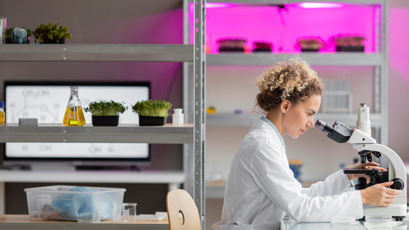
Molecular cytogenetics encompasses all aspects of chromosomal biology, such as the structural and functional organisation of the chromosome and nucleus, chromosomal variations and aberrations and the applications of chromosomal biology in medicine and cancer genetics.
Cytogenetics plays an important role in identifying the genetic predisposition to disease and also in a person’s response to a particular drug.
Molecular biology techniques have evolved from simple tools such as in situ hybridization (ISH), which detects the presence, quantity and location of DNA / RNA sequences in cells and tissues, using a labelled sequence fragment called a probe . These screening tests analyse complicated genomic interactions and help determine the course of a disease.
The growing incidence of cancer and genetic abnormalities, the need for rapid diagnosis of diseases through genetic techniques, as well as the increase in funding (both public and private) leading to the introduction of highly advanced cytogenetic techniques and their wider applications are some of the key factors that are poised to contribute to future market growth.
A growing level of awareness of genetic diseases and an expansion of the pool of research laboratories specialising in molecular biology techniques would be useful for cytogenetic solutions.
Market set to grow in the US and China
The US market is estimated to reach $ 794.6 million by 2022, while China is expected to reach $ 159.7 million by 2026.
The molecular cytogenetics market in the United States is estimated to be worth $ 794.6 million by 2022. China, the world’s second largest economy, is expected to reach an expected market size of $ 159.7 million by 2026, with a CAGR of 8.9% over the analysis period.
Other notable geographic markets include Japan and Canada, each of which will grow 6.3% and 6.8% respectively over the analysis period. In Europe, Germany is expected to grow at around 7.2% CAGR. Software and services segment expected to reach $451.8 million by 2026
Some of the key players in the global molecular biology industry are:
- Abbott Molecular
- Applied Spectral Imaging
- Agilent Technologies, Inc.
- Biological Industries
- Bio-Rad Laboratories, Inc.
- Cytognomix, Inc.
- CytoTest, Inc.
- F. Hoffmann-La Roche Ltd.
- Illumina, Inc.
- Leica Biosystems Nussloch GmbH
- Oxford Gene Technology
- PerkinElmer, Inc.
- SciGene Corporation
- Thermo Fisher Scientific, Inc.
Developments in molecular cytogenetics
Artificial intelligence company based in the UK, DeepMind, has announced for the first time that it has developed a method to accurately predict the structure of folded proteins by the end of 2020 and, by mid-2021, that it has revealed 98.5% of the proteins used in the human body.
DeepMind, an artificial intelligence company, has transformed biology by predicting the structure of nearly every protein known to science in just 18 months, a breakthrough that will accelerate drug discovery and revolutionise basic research
DeepMind has predicted the structure of nearly every protein known to science to date and, using an artificial intelligence called AlphaFold, has solved one of biology’s great challenges in just 18 months. The researchers say the work has already led to advances in the fight against malaria, antibiotic resistance and plastic waste, and could accelerate the discovery of new drugs.
Determining the crumpled shapes of proteins from their amino acid sequences has been a persistent problem in biology for decades. Some of these amino acids are attracted to others, others are repelled by water and the chains form complex shapes that are difficult to pin down.
Today the company announced that it has published the structures of over 200 million proteins – almost all of them listed in the world-renowned archive for protein research, UniProt.
DeepMind has collaborated with the European Institute of Bioinformatics (EMBL-EBI) of the European Molecular Biology Laboratory to create a searchable archive of all this information that is easily and freely accessible to researchers around the world. Ewan Birney of EMBL-EBI calls the AlphaFold Protein Structure Database “a gift to humanity”.
“As someone who’s been in genomics and computational biology since the 1990s, I’ve seen many of these moments come where you can sense the landscape shifting under you and the provision of new resources, and this has been one of the fastest,” he says. “I mean, two years ago, we just simply did not realise that this was feasible.”
Keith Willison of Imperial College London says AlphaFold has undeniably changed the world of biological research, but the problems still need to be solved in protein folding.
“As soon as AlphaFold came out it was wonderful. You just take your favourite proteins and look them up now rather than having to make crystals,” he says. “I did the crystallographic structure of a protein complex, it took me about eight years. People are joking that crystallographers are going to be unemployed.”
Speaking of their progress, Pushmeet Kohli, DeepMind’s scientific team lead, says that the company isn’t done with proteins yet and is working to improve the accuracy and capabilities of AlphaFold.
“We know the static structure of proteins, but that’s not where the game ends,” he says. ‘We want to understand how these proteins behave, what their dynamics are, how they interact with other proteins. Then there’s the other area of genomics where we want to understand how the recipe of life translates into which proteins are created, when they are created and the working of a cell.”
Since its introduction in the 1980s, PCR has become a standard tool in biomedical research.
The equipment (thermocyclers) and reagents (thermostable polymerases, oligonucleotides, etc.) needed for polymerase chain reaction (PCR). They are widely available and relatively inexpensive. An advantage of PCR is its extreme sensitivity, allowing the detection and analysis of low abundance DNA.
This is particularly useful when limited amounts of starting material are available or when few copies of the target sequence exist.
Applications of PCR include cloning of known and novel genomic DNA and cDNA sequences, DNA sequencing, construction of mutant or chimeric DNAs, and quantification of mRNA and DNA. PCR is also used in some difference analysis methods.
Most common techniques
In this section, we will cover some basic techniques in molecular biology. These primers would be useful to give you an overview of the areas, but be mindful that these are not enough to perform these procedures in the laboratory.
Plasmid transformation
Plasmid transformation is a mechanism by which we can introduce genes of interest into bacterial cells. To do this, we isolate another plasmid, add the desired gene and reinsert the plasmid into the bacterial cells.
We can isolate the plasmid through various biochemical means, and the plasmids are small enough to isolate entire plasmids. (We have trouble isolating whole pieces of longer DNA, which tends to break into fragments.)
Once we have isolated the plasmid, we can apply restriction enzymes (like EcoRI) to the specific sequences that we want in the plasmid and gene of interest. Restriction enzymes cleave the plasmid and our gene at the cleavage sites; stick ends (ends of DNA with a single strand between two and four bases long) are often left out.
These sticky ends easily join other matching pieces of DNA; This way we can insert the gene of interest into our plasmid or cut other parts of the plasmid. After adding the restriction enzyme, we add DNA ligase to ligate the plasmid (to reform the covalent bonds that hold the DNA together).
Once we have our plasmid ligated, we want to insert it into bacterial cells. Some bacteria are naturally competent and simply accept plasmids from their environment. However, many other bacteria (like E. coli) are not inherently competent and we need to induce competence to get them to accept the plasmid.
There are electrical methods of competence induction which apply a field and essentially open the bacterium to take up the plasmid; Although most bacteria die, some survive and form colonies.
There are also chemical methods to induce competence, such as adding calcium chloride to E. coli, but their mechanism of action is more complicated.
Finally, once we introduce our plasmid into bacterial cells, we can grow them back into colonies. If necessary, we can apply the selection to recombinant plasmids, which will be discussed later in this primer of the technique.
Electrophoresis
It is a process that allows us to separate DNA fragments according to their molecular weight and to visually examine the different sizes that exist, as well as to measure the size of DNA fragments.
To perform DNA electrophoresis, we start by preparing the DNA fragments of interest. In some cases these come from restriction enzymes applied to larger fragments, but this is by no means required or a feature of electrophoresis.
An agar gel is then produced which is placed in a plate; this plate must have two electrodes on the sides. Once the plate is prepared, we place the DNA in wells in the agar gel on the side of the negative electrode. An electric field is applied between the two electrodes and due to the negative charge of the phosphates, the DNA migrates towards the positive electrode.
The electric field is maintained for a while and then turned off. Smaller fragments encounter less resistance from the gel and therefore move faster, while larger fragments, which have more difficulty passing through the pores of the gel, move more slowly.
The distance travelled depends more or less logarithmically on the size of the molecule, since smaller pieces experience exponentially less resistance than larger ones.Note that with fragments of similar size, it may be impossible to tell them apart because they are placed next to each other. However, you can increase the freeze run time to reduce this.
To visualise the results, we add ethidium to the gel (or after). Ethidium is a dye that binds to DNA and is found in the DNA helix. As DNA migrates, it collects ethidium and therefore has it in much higher concentrations than the surrounding gel. Ethidium glows under ultraviolet light, allowing us to see where the DNA bands are.
Restriction on digestion
A restriction enzyme is a protein that splits DNA into specific sequences. The resulting break in the DNA can be used to insert other pieces of DNA into that location, after which the DNA ligase can be used to re-link the DNA together.
The sequences on which the restriction enzymes act are generally the same from 5 ‘to 3’ (they are DNA palindromes). This is because restriction enzymes are usually dimers with each monomer in the dimer acting on a single strand. If the sequence is palindromic, both monomers will bind in the same position on the strand.
Restriction enzymes are specific to the sequence and also to the type of cut they make. They can create blunt ends in the cut (where the cut is in the middle of the string), resulting in fully double stranded DNA until it breaks. They can also create so-called “sticky tips”. A sticky end is a piece of DNA that has a small single-stranded portion at the end of a strand, which is created because the restriction enzyme cuts the sequence off-centre.
This single-stranded portion can be between two and four bases in length and mates with the single-stranded portion on the other half of the break. Because they fit together, it’s energetically favourable for them to connect (since it forms hydrogen bonds between the single-stranded parts), so it’s more likely to happen; For this reason, these are called “sticky” ends.
When DNA rejoins with hydrogen bonds, there is no reason for it to rejoin in exactly the same configuration as before. For this reason, we can cleave DNA and insert other pieces of DNA (with matching sticky ends) into this region. After that, we can use DNA ligase to covalently link the DNA pieces.
Binding
DNA ligase is an enzyme responsible for the covalent bonding of pieces of DNA or nucleotides. In in vivo DNA replication, the ligase fills in the cuts (unbound pieces of DNA) left behind after the stranded replication delay. (Remember that RNA primers are broken down, so DNA polymerase adds bases to fill the void left by the primer, but cannot covalently attach the new bases to the bases that were there after the primer; this is what DNA ligase does. .)
We can use DNA ligase in combination with restriction enzymes to cut the DNA and then reassemble it. For example, we can use it to create recombinant plasmids.
Ligase works best with the sticky ends left behind by the restriction enzymes, but it also works well with blunt ends, although higher concentrations are required.
Sanger sequencing
Sanger sequencing is a technique for obtaining the sequence of bases (Aa, Cs, Gs and Ts) that make up a piece of DNA. It was one of the first methods developed and, with minor improvements, is still used today. Although it has a much lower throughput than modern methods, it can sequence much larger contiguous sequences (unlike shotgun sequencing methods).
We will start by setting up a modified DNA synthesis reaction. In addition to adding a template strand and a primer (which determines what we sequence), we add sufficient amounts of deoxyribonucleotides (dNTPs). However, we also add a small amount of dideoxyribonucleotides (ddNTPs) to the mixture; the ratio of ddNTP to dNTP is very small, on the order of one in a hundred.
Quantitative PCR (qPCR)
Quantitative PCR (qPCR) is an application of PCR where instead of amplifying the target sequence, you want to measure the concentration or relative concentration of the target sequence in the starting sample.
PCR begins and proceeds as in a conventional reaction. However, the concentration of the target sequence is measured after each iteration of the PCR.
A common way to do this is to have a fluorophore that binds to double-stranded DNA; then, when the DNA cools and reverts to its double-stranded form, the dye binds to it and shows fluorescence. The more DNA there is, the more dye binds and therefore the greater the fluorescence. This allows us to measure and quantify the levels of DNA present after each iteration.
Reverse transcription
Reverse transcription is a process by which we can convert RNA strands back into DNA. The generated DNA is known as complementary DNA (cDNA) and is produced by enzymes known as reverse transcriptase.
The process is started with an oligo (which acts as a primer), reverse transcriptase and nucleotides (dNTP). Transcriptase synthesize a DNA strand complementary to the given RNA sequences. Then the RNA is removed, leaving a strand of DNA with the RNA primer at the beginning. Then, DNA polymerase synthesises a complementary strand to the strand made up entirely of DNA, creating a copy of the original RNA as a strand of DNA.
Note that due to the complexity of this process (multiple polymerases involved, etc.), it turns out to be incredibly error-prone and causes a rather high mutation rate. For example, retroviruses have a genome composed entirely of RNA and use reverse transcription to reproduce; this results in a very high mutation rate between them.